
MR Ergometer Pedal
Medication free, load adjusted patient activation during MR scans
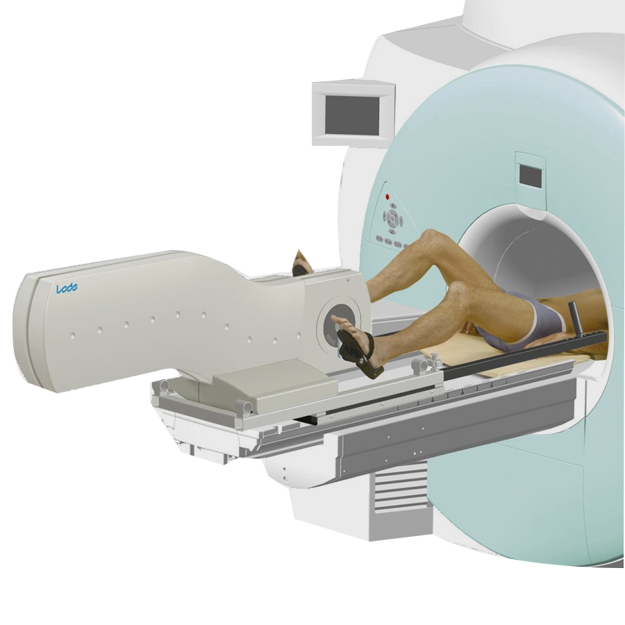
The MR ergometer is an ergometer for use during MR studies. The MR ergometers workload is controlled with an electronic braking principle especially designed for use in a MR environment. The moment of inertia is 8,4 kgm2. The MR ergometer is standard supplied with a control unit and power unit. The standard control unit offers the possibility to read out various parameters like workload, rpm, torque, timer and distance. The power unit is completed with a safety cable for wall fixation. The MR ergometer can be used for MRI scanners up to 3 Tesla.
 A unique ergometer


Overview
Highlights
Unique ergometer for MRI
With this unique ergometer it is possible to perform an exercise test in an MRI. An ergometer on the tabletop of an MRI ensures minimal time stress induction and imaging without coursing artifacts on the imaging.
High standards
Lode is a socially and environmentally responsible company. All Lode products are RoHS/WEE compliant and Lode is ISO 9001:2015, ISO and 13485:2016 certified. All medical products comply to MDD 93/42/EEC, incl. IEC 60601-1.
Compatible with various MRIs at 1.5 and 3 Tesla
Compatible with
– Philips
– Siemens
– GE
– Philips
– Siemens
– GE
Tesla independent
The choice of materials and the special design makes that the Lode MRI ergometer is useable for various Tesla MRIs without giving artifacts on the imaging.
Exercise instead of medicine
When a test subject is able to do exercise, it is always recommended above pharmacologic stress. It allows objective measurement in either level of cardiac conditioning and/or level of cardiac work. It is safe and perfectly reproducible.
Features
Extreme low start up load
The extreme low start-up load of 7 watts and the adjustability in small steps of 1 watt make this ergometer perfectly suitable for many different applications. The standard control unit shows multiple ergometry parameters and you can determine your specific default setting and start-up menu.
GE MRI compatible
The Lode MR Ergometer is compatible with GE Signa and Discovery scanners.
Siemens MRI compatibility
The MRI Ergometer can be used in combination with many Siemens Magnetom MRI scanners, like Skyra, Aera, Verio, Essenza, Prisma, Avanto Fit, Vida, Sola, Altea, Lumina and Spectra.
Philips MRI compatible
The ergometer is compatible with various Philips MRI devices
like Philips Achieva and Ingenia.
like Philips Achieva and Ingenia.
Additional features with PCU
Besides the possibility to program 24 protocols easily, this control unit offers the following features:
– better monitoring because of the additional and larger display
– a perfect combination with BPM
– possibility to measure SpO2
– better monitoring because of the additional and larger display
– a perfect combination with BPM
– possibility to measure SpO2
Low noise
Due to accurate manufacturing and the careful choice of materials the product has an extremely low noise level.
Accurate over a long period of time
The Lode ergometers are supplied with an electro-magnetic braking mechanism of Lanooy (eddy current). The biggest advantage of this braking system compared to a friction braking system is the absolute accuracy and the accuracy over time. Moreover, friction braking systems have more wearing parts.
Small adjustment steps
The workload of the Lode ergometers is adjustable in steps of only 1 watt. Depending on your wishes, the test operator or the test subject can adjust the workload. The steps of 1 watt are possible in the manual mode as well as within protocols.
Service friendly ergometer
Lode ergometers are very service friendly. In general, total costs for spare parts are so low that they are negligible. Furthermore, most options are so easy to install and firmware is so easy to update that labor costs are minimal. Moreover, the ergometer can be cleaned easily.
Up till 3 Tesla
Virtually no interferentie up till 3 Tesla through smart constructions and material use
Accessories
Specifications
*Specifications are subject to change without notice.
Science
Science
- Accelerated cardiac MR stress perfusion with radial sampling after physical exercise with an MR-compatible supine bicycle ergometer.
- An MR-Compatible Treadmill for Exercise Stress Cardiac Magnetic Resonance Imaging
- Arm exercise as an alternative to pharmacologic stress testing: arm exercise stress testing and outcome
- Assessing exercise cardiac reserve using real-time cardiovascular magnetic resonance
- Assessing vascular response to exercise using a combination of real-time spiral phase contrast MR and noninvasive blood pressure measurements
- Atrial volume and function during exercise in health and disease
- Automatic segmentation propagation of the aorta in real-time phase contrast MRI using nonrigid registration.
- Biventricular response to supine physical exercise in young adults assessed with ultrafast magnetic resonance imaging
- Calf Muscle Perfusion at Peak Exercise in Peripheral Arterial Disease: Measurement by First-Pass Contrast-Enhanced Magnetic Resonance Imaging
- Cardiac MRI: a new gold standard for ventricular volume quantification during high-intensity exercise
- Characterizing blood oxygen level-dependent (BOLD) response following in-magnet quadriceps exercise
- Design and testing of an MRI-compatible cycle ergometer for non-invasive cardiac assessments during exercise
- Determinants of pulmonary perfusion measured by electrical impedance tomography
- Dynamic bicycle exercise to assess cardiac output at multiple exercise levels during magnetic resonance imaging
- Effect of Fontan geometry on exercise haemodynamics and its potential implications
- Effect of respiration on cardiac filling at rest and during exercise in Fontan patients: A clinical and computational modeling study
- Effects of Exercise and Respiration on Blood Flow in Total Cavopulmonary Connection
- Exercise capacity in single-ventricle patients after Fontan correlates with haemodynamic energy loss in TCPC
- Exercise stress CMR reveals reduced aortic distensibility and impaired right-ventricular adaptation to exercise in patients with repaired tetralogy of Fallot
- Exercise stroke volume and heart rate response differ in right and left heart failure.
- Exercise testing to estimate survival in pulmonary hypertension
- Feasibility and Reproducibility of Biventricular Volumetric Assessment of Cardiac Function During Exercise Using Real-Time Radial k-t SENSE Magnetic Resonance Imaging
- Flow during exercise in the total cavopulmonary connection measured by magnetic resonance velocity mapping.
- Heart Journal
- High-resolution phase-contrast MRI of aortic and pulmonary blood flow during rest and physical exercise using a MRI compatible bicycle ergometer.
- Improving the physiological realism of experimental models.
- Interaction between respiration and right versus left ventricular volumes at rest and during exercise: a real-time cardiac magnetic resonance study
- Leg lean mass correlates with exercise systemic output in young Fontan patients.
- Magnetic Resonance Augmented Cardiopulmonary Exercise Testing: Comprehensively Assessing Exercise Intolerance in Children with Cardiovascular Disease
- Measurement of aortic and pulmonary flow with MRI at rest and during physical exercise.
- MR augmented cardiopulmonary exercise testing-a novel approach to assessing cardiovascular function.
- MRI evaluation of left ventricular volume in children and adults during in-scanner exercise.
- Paradoxical Increase in Ventricular Torsion and Systolic Torsion Rate in Type I Diabetic Patients Under Tight Glycemic Control FREE
- Peripheral arterial disease assessment: wall, perfusion, and spectroscopy
- Quantitative abdominal aortic flow measurements at controlled levels of ergometer exercise
- Respiratory Effects on Fontan Circulation during Rest and Exercise Utilizing Real Time Cardiac Magnetic Resonance Imaging
- Stroke volume response during exercise measured by acetylene uptake and MRI
- The effect of supine exercise on the distribution of regional pulmonary blood flow measured using proton MRI
- The response of the pulmonary circulation and right ventricle to exercise: exercise-induced right ventricular dysfunction and structural remodeling in endurance athletes
- UNDERSTANDING THE CARDIOPULMONARY CIRCULATION IN SICKLE CELL DISEASE: USING AN MR AUGMENTED CARDIOPULMONARY EXERCISE TESTING TECHNIQUE
Support
Service
Part number
937901
Interfacing
Interfacing protocols
AEI Technologies
AMEDTEC Medizintechnik Aue GmbH
BTL
CareFusion
Cognimed
Cortex
Custo Med
Delmar Reynolds
Edan Instruments Inc.
Enverdis GmbH
ET Medical
GE Healthcare
Korr Medical Technologies Inc
Labtech
Lode
Med-tronik
Medical Graphics
Mesa Medizintechnik
Mortara
Nihon Kohden
Norav
nSpire Health
Parvomedics
Philips Medical
Progetti
Pulse Biomedical, Inc.
Quinton
Schiller
Seiva
Welch Allyn
1350K
1500 K / 1550K – Cardiofax V
600 USB system
9320K
AT-10
AT-104 PC
AT-110
AT10 Plus
BreezeSuite Software
BTL-08 ECG
Cardiax PC-EKG
Cardio Coach Monitor
Cardio Collect 12
Cardiologic Explorer 12C Ergo
CardioPerfect
Cardiosoft
Cardiosoft + Corina
Cardiosoft MAC 1200 ST
CS200
Cubestress
Custo Diagnostic Stress ECG
EC12 R/S
ECG Pro
Edan EC-12 EXPRESS
Edan ST-1212 STRESS ECG
Ergolog
GE Case
GE Case 8000
GE Case Basic
GE Case Performance
GE Case Premium
GE Case Value
Lactware Software
Lode Ergometry Manager
MAC 5000
MAC 5500st
MasterScreen® ECG / Viasys Oxycon Pro
Metasoft
MOXUS Modular Metabolic System
Multi Control
MyStress system
PC ECG 1200
Q-Stress
QRS-card
StressVue
TM Cardio 1000/2000
TrueOne
X-scribe
XR-200
Reviews
There are no reviews yet.